-
[ July 13, 2012]
STED Super-resolution Optical Microscopy realized
-
Recently, Dr. Peng Xi, associate professor in Department of Biomedical Engineering, and his team have demonstrated a CW Stimulated Emission Depletion (STED) microscope using a Ti:Sapphire oscillator. Taking advantage of this new type of microscopy, they resolved three cytoskeletal elements (microtubules, intermediate filaments, and actin filaments). The results have been published on PLoS ONE.
Fluorescence microscopes with the most advanced confocal geometries available yield optical resolution approaching the theoretical Abbe diffraction limit of ~200 nm, but this is still larger than many subcellular structures, which are too small to be resolved in detail. Bypassing the optical diffraction limit in far-field optical microscopy has been realized by three key pioneering technologies: STED microscopy, Saturated Structured Illumination Microscopy (SSIM) and STochastic Optical Reconstruction Microscopy (STORM)/Photo-Activated Localization Microscopy (PALM). STED microscopy was the first and most direct approach to overcoming the diffraction limit for far-field nanoscopy. STED microscopy uses an intense doughnut-shaped beam surrounding the excitation focus to switch the fluorophore(s) in the sample to a ‘‘dark’’ state through stimulated emission. This effectively eliminates the periphery of the Point Spread Function (PSF), resulting in a narrower PSF, or super-resolution. Scanning a sharpened spot through the specimen renders images with subdiffraction resolution.
In spite of the major advantages, the alignments in a STED system is typically complicated comparing to the other techniques because the two beams must be overlapped at nanometer level precision. Leica corp. has launched its commercial STED systems, but these are prohibitively expensive for most users. On the other hand, the Ti:Sapphire laser which is widely used in two-photon microscopic system has the possibility to expand its application to STED nanoscopy. Basing on this point, Xi’s group tuned a 6W pump Ti:Sapphire oscillators as STED beam, and achieved 60 nm imaging which is one tenth of flourescent wavelength.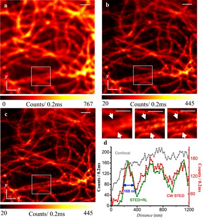
Fig. 1 Comparison of confocal and STED super-resolution nanoscopy on keratin imaging.This work is the first time to realize STED super resolution microscopic imaging in China. Thanks to the widely available Ti:Sapphire oscillators in multi-photon imaging system, this work suggests easier access to setup super-resolution microscope via the synchronization-free STED.
Dr. Peng Xi is dedicated to the research of biomedical optics, and his research interest includes: STED super-resolution microscopy, confocal laser scanning microscopy, multi-photon microscopy, and optical coherence tomography (OCT).
Some of the recent publications and media reports of Xi group are listed below:
1. Achieving λ/10 Resolution CW STED Nanoscopy with a Ti:Sapphire Oscillator
2. Sciencenet.cn Researchers realized STED super-resolution nanoscopy
3. Laser oblique scanning optical microscopy (LOSOM) for phase relief imaging
4. CRAFT: Multimodality confocal skin imaging for early cancer diagnosis
5. OCTNews.org has reported their OCT work on Feature of the Week.