-
[ February 24, 2014]
Progress on phase-sensitive microscopy made by Dr. Peng Xi’s group
-
Recently, a new progress on phase-sensitive microscopy has been made by Dr. Peng Xi’s group from Department of Biomedical Engineering (BME), College of Engineering at Peking University and his collaborators. On the base of confocal microscopy, a new phase contrast imaging mechanism, termed Schlieren-confocal microscopy has been developed. This work is recently published in Optics Letters (http://www.opticsinfobase.org/ol/abstract.cfm?uri=ol-39-5-1238 ).
Phase objects are objects with little light absorption. In spite of their common existence in biomedical research, observation of phase objects is still a challenge. Traditional techniques using prisms, polarizer, phase plates or spatial light modulator are either too complicated or unable to be directly adapted to confocal microscope systems, which are widely used in biological research. Prof. Peng Xi's group developed a new low-cost phase imaging microscopy totally compatible with confocal systems. A quantitative analysis derived from Fraunhofer diffraction theory reveals a good linearity and sensitivity between image intensity and sample phase gradient, indicating its potential as a new microscopy technique for multi-mode imaging. This method has already been successfully applied to the imaging of mouse kidney slides as well as HeLa cells. It is another breakthrough after their previous work Laser Oblique Scanning Oblique Microscopy (LOSOM) on Optics Express and Scientific Reports.
(link: http://www.opticsinfobase.org/abstract.cfm?uri=oe-20-13-14100
http://www.nature.com/srep/2013/130503/srep01762/full/srep01762.html)
The first author of this article is Mr. Hao Xie, a PhD student in Dept. BME, and the corresponding author is Dr. Peng Xi. This work is supported by grants from the National Instrumentation Program, the "973" Program of China, and the National Natural Science Foundation of China.
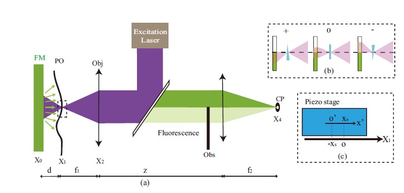
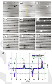
Figure 1: Schematic diagram and result of Schlieren confocal microscopy.