-
[ February 22, 2015]
Breakthrough in the research of super-resolution fluorescent protein development
-
The invention of Green Fluorescent Protein (GFP) has been awarded the 2008 Nobel Prize in Chemistry, for its capability of labeling the specific target gene position through gene modification. Further, the gene modified light-activated fluorescent protein (PA-FP) offers single molecule property, leading to the realization of the super-resolution microscopy, which won the 2014 Nobel Prize in Chemistry. Since then, the development of super-resolution microscopy has been dedicated on the high spatio- temporal resolution of visualizing dynamic live cell subcellular organelles. Of the various techniques, super-resolution optical fluctuation imaging (SOFI) can break the single-molecule emitting limit, therefore it quickly becomes a hot research topic as it offers high speed super-resolution imaging by allowing multiple fluorescent molecules to be at ON state simultaneously.
Recently, Dr. Peng Xi’s group from the College of Engineering, Peking University, Dr. Pingyong Xu’s group from Institute of Biophysics, Chinese Academy of Sciences, and Dr. Jianxin Peng’s group from Central China Normal University, jointly developed a novel fluorescent protein suitable for high speed live cell super-resolution fluorescence microscopy, and named it Skylan-S. The notable features of Skylan-S include: a 6-8 times higher intensity and 4-fold larger fluctuation range than Dronpa, a standard fluorescent protein used in super-resolution microscopy. Its photostability and switching cycles are also systematically improved. Meanwhile, as Skylan-S is a monomer, it is compatible for live cell imaging. Based on these characteristics, it has been applied to SOFI and PALM super-resolution imaging. In SOFI imaging, 1.5 seconds high spatiotemporal resolution imaging (3 milliseconds per frame, 500 were reconstructed SOFI) has been achieved in imaging the ring structure of CCP with 4th order SOFI, and the spatial resolution was better than 100 nm. Meanwhile, Skylan-S is able to dynamically observe subcellular structures for more than 60 seconds, while Dronpa cannot resolve the ring structure even in the first frame, and more than half the dye is photobleached in 30 seconds. Further, the imaging of Skylan-S and Dronpa labeled cells actin filaments show that, Skylan-S can show the enrich grayscale to display the fine structure of different concentrations of proteins, while Dronpa can only provide very limited dynamic range with poor SNR. Skylan-S also demonstrates high single molecule property for PALM imaging. Moreover, the high brightness and photostability also enable its wide application in traditional confocal and two-photon imaging.
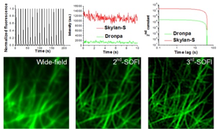
Fig. 1 Skylan-S has greater fluorescence intensity and fluctuation range than Dronpa, making it more suitable for high-order SOFI super-resolution imaging.
The co-first authors of this work are Xi Zhang (Inst. Biophysics, CAS) and Xuanze Chen (PKU). This work is recently published in ACS Nano, an internationally renowned research journal. The work is funded by the national "973" program, the National Special Instrument Development Projects, the National Natural Science Foundation of China, Beijing Natural Science Foundation of China, and Chinese Academy of Sciences.Dr. Peng Xi’s group at Peking University is dedicated on super-resolution microscopy techniques. Several research work have been published since 2015: through inverse multiplexing of spectral separation they achieved joint tagging SOFI, which is published by Nature Publishing Group’s Scientific Reports, and three-dimensional super-resolution microscopy has been demonstrated through the application of spinning disk confocal with different super-resolution modalities (3D-MUSIC), which is published by Nano Research, a journal jointly published by Springer and Tsinghua University Press.
Original link:
http://pubs.acs.org/doi/pdf/10.1021/nn5064387